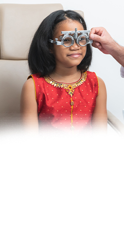
Protect & Preserve Your Sight from Eye Diseases
At Seaton Vision Care, we understand vision is an important part of many families’ everyday lives. But we also understand that there’s more to sight than just clarity and wearing a stylish pair of glasses—your eye health is just as important, if not more so.
Early detection and timely management of eye disease are important for preserving your vision. Through modern technology and our personalized approach, you can gain a deeper understanding and appreciation of your eyes, how they work, and what you can do to support them.
Whether it’s a concern like pink eye or something more serious like glaucoma, we’re here to support your family every step of the way. Book your next eye exam at Seaton Vision Care today.
Book Appointment
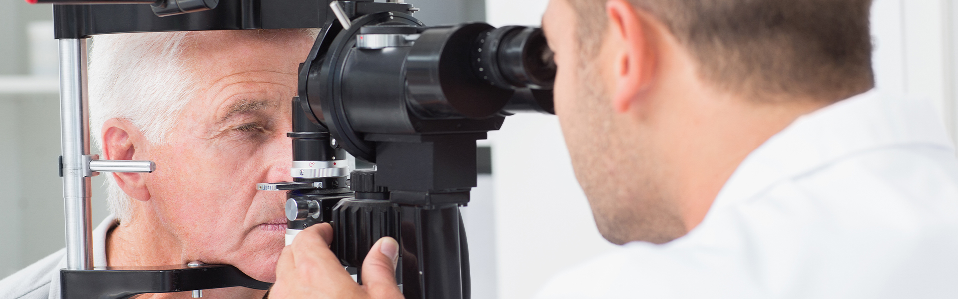
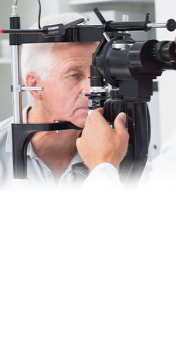
The Importance of Dilated Eye Exams
Dilated eye exams are an invaluable tool for assessing eye health. Dilating your eyes helps us view the structures inside them, making it possible to diagnose conditions that may otherwise go unnoticed.
By having you come in for regular dilated eye exams, we aim to catch potential issues early, opening the opportunity to preserve your eye health with personalized care and modern techniques.
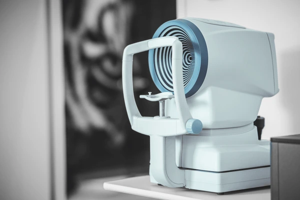
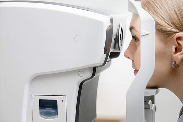
Innovative Technology for Early Diagnosis
We place great importance on giving you a clear understanding of the technology that supports our personalized approach.
Many eye diseases can develop without symptoms in the early stages. However, earlier detection and intervention can lead to a much better diagnosis. The only way to do this is by regular, comprehensive eye health exams with a dilated retinal examination.
We want to help you feel confident in knowing you have comprehensive support for your eyes, and that starts by helping you learn all about the innovative techniques and technology we use for eye exams.
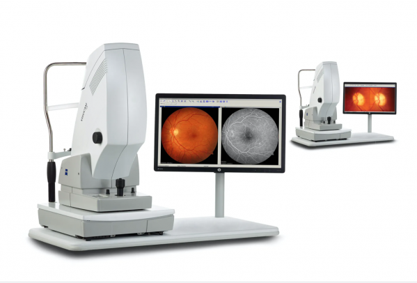
Fundus Photography
Fundus photography is a technique that involves taking highly detailed photographs of the back of your eye (known as the fundus). It helps us track changes over time, identify abnormalities in your retina, and recommend appropriate treatments.
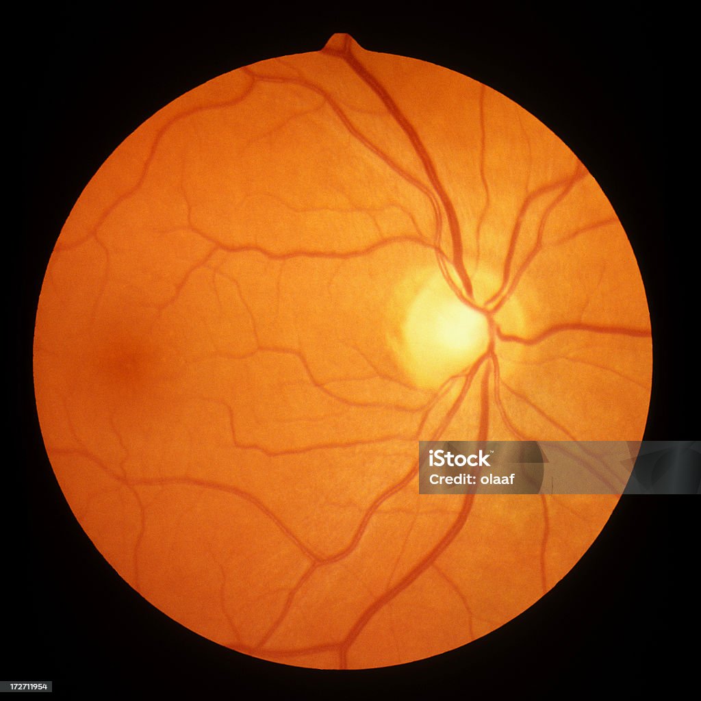
With these images, we can find blockages in blood vessels, bleeding in the retina, signs of early degeneration, genetic diseases, holes in the retina, retinal detachments, tumours, swelling of the optic nerve, and more.
Fundus Autofluorescent (FAF) Imaging
Fundus autofluorescence (FAF) imaging is a diagnostic tool that helps us see deeper into your eyes. FAF imaging uses a special camera to capture a map of natural fluorescent molecules within your retina. These molecules can reveal changes in the health and function of your retinal cells.
FAF Imaging is a valuable tool for diagnosing and monitoring a variety of eye conditions, including:
- Age-related macular degeneration (AMD)
- Diabetic retinopathy
- Inherited retinal diseases
- Macular telangiectasia (MacTel)
- Central serous chorioretinopathy (CSC)
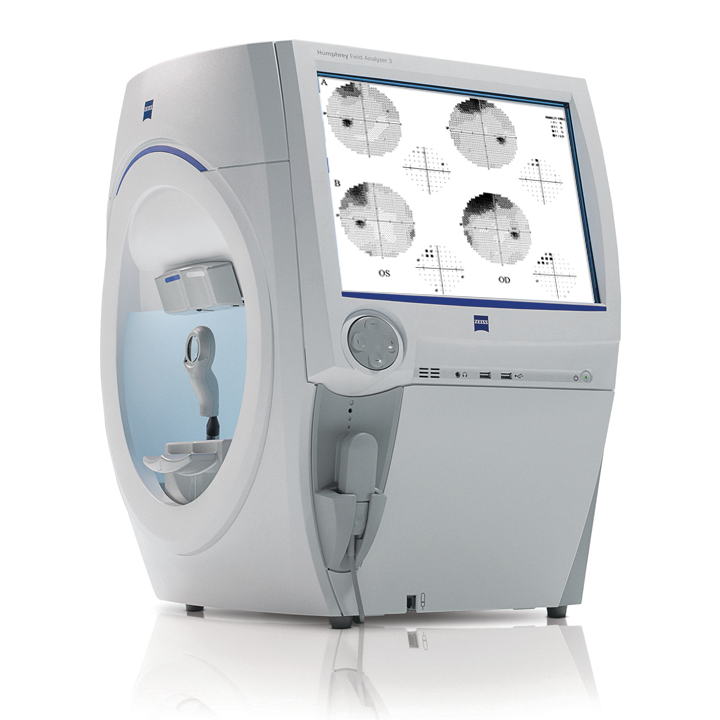
Visual Field Testing
Visual field testing, specifically with the Zeiss Humphrey Field Analyzer, is a painless computer-aided test that maps your peripheral vision or side vision.
During the test, you will sit comfortably and stare at a central target light. Lights of varying intensity will appear at different locations in your field of vision. Your job is to press a button whenever you see one of these lights.
The results of the visual field test are presented as a map showing areas where you may have lost some vision. This information can be crucial in diagnosing and monitoring a variety of eye conditions, including:
- Glaucoma
- Brain tumours or strokes
- Macular degeneration
- Other retinal diseases
Corneal Topographer
A corneal topographer is a an instrument that creates a detailed, three-dimensional map of the curvature of your cornea, the clear dome-shaped surface at the front of your eye. This painless test measures the shape of your cornea to assess its overall health and identify any irregularities.
Corneal topography is a valuable tool for diagnosing and monitoring a variety of corneal conditions, including:
- Keratoconus
- Astigmatism
- Corneal injuries
- Corneal ectasia
By providing a precise picture of your corneal shape, corneal topography allows your us to diagnose potential problems early and recommend appropriate treatment options.
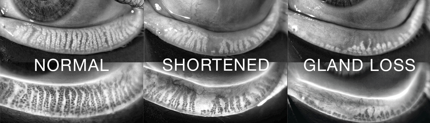
Meibography
Meibography is an imaging test that allows your eye doctor to examine your meibomian glands. These tiny glands, located in your eyelids, produce oils that form an important layer of your tear film. A healthy tear film keeps your eyes lubricated and comfortable and helps prevent infections.
Meibography helps your doctor assess the health and function of your meibomian glands.
Meibography can help diagnose a variety of conditions affecting the tear film and eyelids, including:
By identifying these conditions early, we can recommend treatments to improve your tear film function and alleviate symptoms like dry eye and irritation.
Optical Coherence Tomography (OCT)
OCT testing is a noninvasive imaging technique that provides high-resolution, cross-sectional images of the retina, revealing subtle changes that can indicate early signs of various eye conditions, such as glaucoma, age-related macular degeneration, and diabetic retinopathy.
OCT testing requires dilation of the pupils but does not require a needle in the arm and doesn’t involve touching the eye.
If you’re interested in learning more, click here for more information.
Common Eye Diseases & Conditions
At Seaton Vision Care, we’re focused on the early detection, diagnosis, and management of various eye diseases and conditions. Through regular eye exams, we can monitor your eyes and identify early signs of several conditions, potentially preventing further damage.
Recognizing common eye problems early can be the difference between making minor adjustments to your treatment and the need for emergency care.
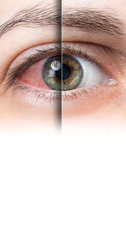
Red Eyes & Infections
Several conditions may lead to red eyes and eye infections, including conjunctivitis (pink eye), dry eye, uveitis, and allergies.
Not all red eyes are pink eye or an infection. Visit our team first to learn more about an effective treatment plan that can help you address red eyes and infections.
Our mission is to quickly identify the underlying cause of redness to provide essential guidance on preserving your eye health, vision quality, and comfort. Please visit our emergency eye care page to learn more about how we can address concerns like sudden redness and eye irritation.
Keratoconus
Keratoconus is a progressive condition that affects the cornea. It may cause distorted vision, light sensitivity, and possible eye discomfort. At Seaton Vision Care, we can diagnose this issue and refer you to specialists for treatment strategies like scleral lenses.
For keratoconus care, we don’t just refer—we also:
- Confirm the diagnosis using our in-house corneal topographer
- Refer out for corneal cross-linking with our affiliated corneal specialists
- Provide follow-up care after your corneal cross-linking procedure
- Help correct your vision after surgery with corrective glasses, soft contacts, or refer out for scleral lenses.
Cataracts
Cataracts are a common eye condition that can affect your vision. They occur when the eye’s lens becomes cloudy, leading to blurred or hazy vision. Adults over the age of 60 and people with diabetes may be at a higher risk of developing cataracts.
Cataracts can be removed with a surgical procedure known as cataract surgery.
Glaucoma
Glaucoma is a group of eye diseases that affect the optic nerve. They can cause permanent vision loss over time and can often develop without any symptoms.
High internal eye pressure is a common identifier for glaucoma, but it can also develop when your eye pressure is within normal limits. Glaucoma is also more common if you have a family history of the disease.
Getting routine eye exams is crucial for detecting glaucoma early—before it causes vision loss.
Age-Related Macular Degeneration (AMD)
AMD affects a small area at the retina’s center called the macula, leading to a loss of central vision. This disease typically develops in adults 55 and older, making it difficult to read, drive, or even recognize faces.
Diabetic Eye Diseases
People with diabetes can be at a higher risk of developing certain eye conditions because of damage to blood vessels in the eye. Regular eye exams are crucial for early detection and management of conditions like diabetic retinopathy.
To learn more about these issues, please visit our diabetic eye exam page.
Book Your Next Eye Exam Today
Consistent eye health exams, including dilated eye exams, are instrumental in diagnosing eye diseases in their early stages—before they cause noticeable vision changes. When you visit us, we can help you determine the right eye exam frequency for your family.
Book your family’s next eye exams today and take the first step towards protecting your sight.
Book AppointmentOur Location
Visit our practice near the Duffin Heights neighbourhood in north Pickering. You can find us in the Seaton Centre, the shopping center off Brock Road and Palmers Sawmill Road.
Our Address
- 2460 Brock Rd, Unit C7
- Pickering, ON L1X 0J1
Contact Information
- Phone: 905-428-3330
- Fax: 905-428-2010
- Email: [email protected]
Hours of Operation
- Monday: Closed
- Tuesday: 11:00 AM – 7:00 PM
- Wednesday: 11:00 AM – 6:00 PM
- Thursday: 11:00 AM – 7:00 PM
- Friday: 10:00 AM – 5:00 PM
- Saturday: 10:00 AM – 5:00 PM
- Sunday: Closed
*Closed on Saturday on long weekends
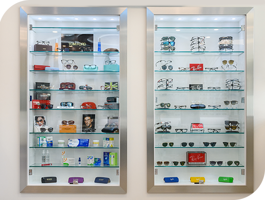
Our Services

Our Brands


Our Google Reviews
-
Seaton Vision Care
- 2460 Brock Rd, Unit C7
- Pickering, ON L1X 0J1
-
P: 905-428-3330
F: 905-428-2010
[email protected]